Sciatica: Going Beyond Common Myths And The Roadmap To Relief

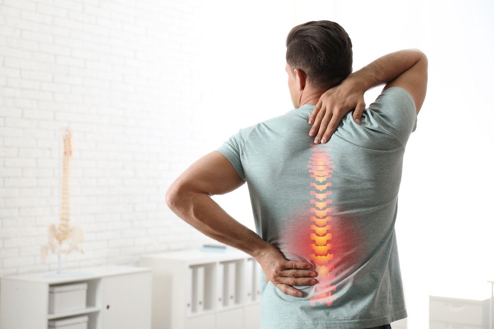
Sciatica – a word which brings a frown to the faces of many thinking it is an incurable entity. The truth is it is one of the most commonly misunderstood causes of pain radiating down from the back and along the leg.

 Many patients believe that Sciatica almost always means a “slipped disc,” that complete bed rest is mandatory, or that surgery is unavoidable.

As   Physiatrists (specialists in Physical Medicine & Rehabilitation), we often see patients who keep suffering unnecessarily because of these gross misconceptions.

The good news is that most patients with sciatica improve with appropriate conservative treatment, and modern techniques of Pain management & rehabilitation offers multiple effective ways to control pain, restore mobility and prevent relapse.

Debunking Common Myths Regarding Sciatica:

Myth 1: Every pain radiating down the leg is sciatica.

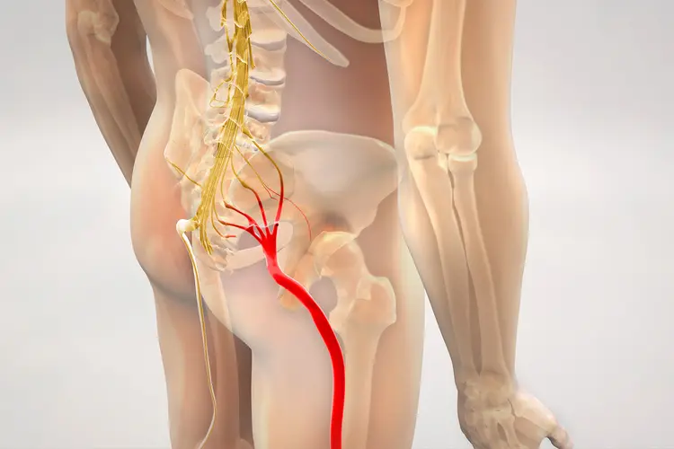
Not necessarily. True sciatica usually involves compression, and subsequent irritation, of the sciatic nerve roots in the lower spine, producing pain that travels from the lower back or buttock into the thigh and leg. Several other conditions like Spinal Canal Stenosis, Spondylolisthesis, Sacroiliac joint dysfunction, Hip joint Osteoarthritis etc can cause similar symptoms. Thus, a thorough clinical assessment by a Physiatrist is a must to avoid misdiagnosis and mismanagement.

Myth 2: Sciatica always means a slipped disc.

No, a disc herniation is definitely common cause, but it is absolutely not the only one. As mentioned earlier, age-related changes such as spinal canal narrowing, bone spurs and degenerative changes can also irritate nerve roots.

Myth 3: Bed rest is the best treatment.

Incorrect. Prolonged bed rest often aggravates muscle weakness, stiffness, decreased confidence in movement, slower and poorer recovery. Unless there is a specific medical reason to restrict activity, staying active within tolerable limits and under guidance of a Physiatrist is generally advisable.

Myth 4: Exercise will worsen the nerve problem.

The correct exercise, prescribed at the right stage, is actually a station on the path to recovery rather than a threat to it. Exercises, as part of supervised Rehabilitation protocols, are advised in accordance with the patient’s symptoms, neurological findings, strength, flexibility and functional requirements.

Treatment is customized to the patient and no one plan fits all.

Myth 5: Sciatica inevitably requires surgery.

This is surely the most important misconception. Most patients can be successfully treated without surgery. Surgery is generally considered when there is significant or progressive neuromuscular deficit like muscle weakness and/or  atrophy,  serious underlying pathology like tumors , or persistent disabling symptoms despite appropriate non-surgical management.

Why and When to Consult a Physiatrist??

A Physiatrist looks beyond treating just the pain. The aim of treatment is to go the depth of the issue as to why the pain occurs, how it affects movement and function, and what can be done to restore normal mobility and ADLs (Activities of Daily Living).

A consultation is particularly useful when pain refuses to disappear completely, returns repetitively, interferes with rest and work, or if it is associated with abnormal sensations of tingling, numbness or muscle weakness.

A Physiatrist carries out a detailed neurological and musculoskeletal examination and determines whether imaging or other investigations are actually necessary.

Most importantly, an MRI abnormality does not always automatically explain someone’s pain. Disc bulges and degenerative changes are more often than not, very common even in many people without symptoms. Management should therefore be based on the patient’s clinical presentation and examinations, rather than an MRI report alone.

Techniques for Pain Relief

Treatment is highly individualized and no one protocol fits all. Depending on the nature , time of  presentation, the occupation of the patient and his/her daily routine, it may include medications for a variable period, physical activity modification, heat and/or other physical modalities, and a structured  rehabilitation programmed, supervised by a Physiatrist.

 Therapeutic exercises and Physical therapy are one of the pillars for the foundations of long-term recovery. Treatment often includes exercises targeted at improving spinal & hip mobility, strengthening of muscles of the core and lower limb, corrections of posture and movement patterns and gradual rebuilding of functional capability.

Patient Education is equally important. Patients should understand which activities amplify their symptoms, how to modify them temporarily and, most importantly, how to gradually return to normal activities rather than becoming apprehensive of movement.

Role of Interventional Pain Management

When pain control remains inadequate despite adequate conservative treatment, interventional pain management may sometimes provide much-needed relief and facilitate progression to rehabilitation.

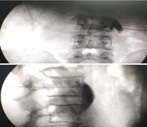
Depending on the diagnosis and clinical findings, procedures such as targeted image -guided (under fluoroscopic or ultrasound guidance) transforaminal epidural or selective nerve-root blocks may be considered. These are not simple “pain-killer injections.” They are meticulously selected well-researched interventions intended to mitigate inflammation and pain around an irritated nerve root, potentially creating a pain-free hiatus during which enables the patient to participate more effectively in rehabilitation.

However, interventional pain management should not be viewed as a substitute for therapeutic exercises and functional restoration. The objective is not merely to make the pain disappear temporarily, but to help the patient move, rehabilitate and return to normal life.

Why Surgery Is Almost Never Required

The natural history of most cases of sciatica is relieving. With time and appropriate non-surgical management, symptoms often improve substantially and mostly disappear. Even when a disc herniation is responsible, the body’s natural healing processes can contribute to improvement, aided by supervised conservative techniques.

Surgery therefore has a selective role but not completely unimportant. It may become necessary in scenarios with severe and/or progressive weakness, significant neuromuscular deficit,  or persistent severe symptoms that hamper with day to day life,  despite an adequate trial of appropriate conservative treatment.

The decision should never ever be based simply on the presence of a “disc bulge” on an MRI.

The Bottom Line

Sciatica should not be feared, ignored or automatically considered as a surgical disease.  A thorough clinical examination, the correct diagnosis, adequate pain control, targeted rehabilitation and gradual return to activity – all under the supervision of a Physiatrist, can help most patients recover without resorting to surgery.

As Physiatrists, our approach is to treat the person as a whole, not just the MRI report or the pain score. If you have pain travelling from your back or buttock towards your leg, especially when accompanied by numbness, tingling or weakness, an early assessment can help identify the cause and create a individualized path to recovery.
Knee Osteoarthritis

Knee Pain: A Comprehensive Guide to Non-Surgical Management

Knee pain is one of the most common health problems seen in adults, especially after the age of 40–50 years.

For the medical management of this condition, a patient should consult a Physiatrist (Physical Medicine and Rehabilitation Specialist). A physiatrist focuses on reducing pain, improving movement, and helping people return to their daily activities without surgery whenever possible.

What is Osteoarthritis of the Knee?

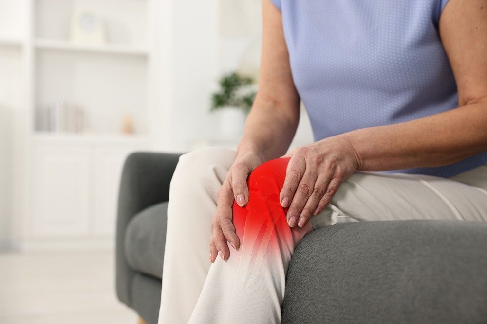
Osteoarthritis (OA) is a condition in which the protective cartilage covering the ends of the bones gradually wears away. As a result, the knee joint becomes painful, stiff, and swollen.

Common Symptoms

  • Pain while walking or climbing stairs
  • Morning stiffness lasting a few minutes
  • Swelling around the knee
  • Difficulty squatting or sitting cross-legged
  • A cracking or grinding sound while moving the knee
  • Reduced ability to walk long distances

OA is not simply a disease of ageing. Excess body weight, previous injuries, genetics, and lifestyle also play important roles.

What Does a Physiatrist Do?

A physiatrist looks beyond the X-ray. Instead of treating only the damaged joint, they evaluate:

  • Pain level
  • Muscle strength
  • Walking pattern
  • Balance
  • Joint movement
  • Daily activities
  • Occupation
  • Lifestyle and fitness

The goal of treatment is to reduce pain, improve function, and delay or even avoid surgery whenever possible.

Treatment Options

1. Patient Education and Counseling

Patients are taught:

  • Safe activities
  • Correct posture
  • Joint protection techniques
  • How to avoid unnecessary stress on the knee

2. Weight Management

Every extra kilogram of body weight places several kilograms of additional force on the knee during walking.

Even losing 5–10% of body weight can significantly reduce pain and improve mobility.

Weight reduction combined with exercise gives the best results.

3. Exercise Therapy

Exercise is the most effective long-term treatment for knee OA.

A trained physiotherapist, under the guidance of a physiatrist, may prescribe:

Strengthening Exercises
  • Straight leg raises
  • Quadriceps strengthening
  • Mini squats
  • Step-ups
Stretching Exercises
  • Reduce stiffness
  • Improve flexibility
Balance Training
Aerobic Exercises
  • Walking
  • Cycling
  • Swimming
  • Water exercises

Regular exercise often reduces pain more effectively than prolonged rest.

4. Pharmacological Management

Treatment may include:

  • NSAIDs
  • DMOADs (Diacerein, Glucosamine, etc.)
  • Nutraceuticals (Chondroitin, Collagen Peptides, Univestin, etc.)
  • Other medications depending on the individual’s condition

Interventional Pain Procedures

When pain continues despite medicines and exercise, a physiatrist may recommend interventions with or without ultrasound (USG) guidance.

Corticosteroid Injection

This injection helps reduce inflammation inside the joint. It is useful when:

  • The knee is swollen
  • Pain suddenly worsens
  • Exercise becomes difficult

Pain relief may last from several weeks to a few months.

Hyaluronic Acid Injection

Hyaluronic acid acts like a lubricant inside the knee. It may:

  • Improve joint movement
  • Reduce pain
  • Help some patients remain active for longer

Results vary from person to person.

Platelet-Rich Plasma (PRP)

PRP is prepared from the patient’s own blood.

Platelets contain growth factors that may help reduce pain and improve knee function in selected patients.

Genicular Nerve Block

Pain signals from the knee travel through small nerves called the genicular nerves.

A local anaesthetic, sometimes combined with corticosteroid, is injected around these nerves to reduce pain.

Radiofrequency Ablation (RFA)

For patients with long-standing knee pain who are not suitable for surgery or wish to postpone it, radiofrequency ablation may be considered.

In this procedure:

  • A special needle is placed near the pain-carrying nerves.
  • Heat generated by radiofrequency energy reduces the nerves’ ability to transmit pain.

Pain relief may last 6–12 months or longer in selected patients.

The procedure does not damage the knee joint—it only interrupts pain signals.

Emerging Treatments

Newer treatments are being studied, including:

  • Bone marrow concentrate
  • Stem cell-based therapies

While some patients report improvement, many of these treatments are still under research, and not all have strong evidence for routine use.

A physiatrist can help determine whether such treatments are appropriate.

Assistive Devices

Sometimes simple devices make a big difference. These include:

  • Walking sticks
  • Knee braces
  • Shoe inserts
  • Appropriate footwear

Using the correct aid reduces stress on the painful knee.

Lifestyle Changes

  • Avoid prolonged squatting.
  • Limit frequent stair climbing when painful.
  • Stay physically active.
  • Take short breaks during long periods of standing.
  • Maintain a healthy weight.
  • Wear comfortable, supportive footwear.

Surgical Management

Surgery may be required only in advanced conditions when all conservative measures fail. A PMR specialist can help assess the need for surgical treatment such as Total Knee Replacement (TKR).

Post-Operative Rehabilitation

Rehabilitation under a physiatrist plays a key role in restoring strength, flexibility, and independence.

Take-Home Message

Osteoarthritis of the knee is a common condition. It does not mean the end of an active life.

With the right combination of:

  • Education
  • Exercise
  • Weight management
  • Medicines
  • Image-guided injections
  • Advanced pain procedures
  • Rehabilitation

Most people can reduce pain, improve mobility, and continue doing the activities they enjoy.

A physiatrist works with each patient to create a personalized treatment plan, helping them move better, stay independent, and improve their quality of life.

Back Pain: A Silent Epidemic Affecting Millions

Back pain has emerged as one of the most common health complaints worldwide, affecting 50% to 84% of the global population at some point in life. In India, nearly 60% of individuals report experiencing back pain, cutting across age groups and lifestyles.

Understanding the Basics

In most cases, back pain stems from minor sprains of muscles or ligaments—often resolving within a week with rest and basic care. However, the body sometimes uses back pain as an alarm to signal more serious underlying issues.

When to Take Back Pain Seriously

Certain symptoms should never be ignored:

  • Pain that worsens at night or interrupts sleep
  • Morning stiffness lasting over 30 minutes
  • Radiating pain to one or both legs
  • Gradual weakness in the limbs
  • History of trauma
  • Associated symptoms like fever, joint swelling, chronic cough, or unexplained weight loss
  • Pain lasting more than a month

These could indicate conditions that require timely medical evaluation.

Common Causes of Back Pain

While most back pain cases are due to simple muscular issues, several conditions may be responsible:

  • Degenerative changes (spondylosis)
  • Inflammatory conditions (spondylitis)
  • Disc problems, such as bulging or herniation
  • Facet joint arthritis
  • Osteoporosis or vertebral fractures
  • Kidney stones, infections, or—in rare cases—cancer metastasis

Interestingly, imaging studies often show degenerative changes in people above 45, yet many remain pain-free. This means scans should always be interpreted alongside clinical symptoms.

The Role of Posture and Nerve Compression

Modern lifestyles involving prolonged sitting, computer use, or driving with incorrect posture place undue stress on the spine. Improper ergonomics can lead to chronic discomfort and, in some cases, disc-related nerve compression—commonly known as sciatica. This may cause pain, numbness, or tingling in the legs.

How Is Back Pain Diagnosed?

An accurate diagnosis begins with a detailed history of the pain’s onset, nature, location, and any radiation to the limbs. This is followed by a physical examination to identify signs of nerve involvement or inflammation.

Initial investigations typically include blood tests and X-rays. Advanced imaging like MRI is reserved for cases where the diagnosis remains unclear or if symptoms suggest nerve or spinal cord involvement.

Treatment Approaches

  1. Medications

Anti-inflammatory drugs, muscle relaxants, and sometimes neuropathic agents like gabapentin are commonly used. Supplements such as calcium or Vitamin D may be prescribed where needed.

  1. Exercise

Once acute pain subsides or the underlying cause is identified, exercises become a crucial part of recovery. Targeted back and core strengthening can prevent recurrence.

  1. Physiotherapy Modalities

Often use tools like TENS (Transcutaneous Electrical Nerve Stimulation) and therapeutic ultrasound, as well as LASER to reduce pain and inflammation.

  1. Image-Guided Injections

For cases resistant to conventional therapy, minimally invasive injections can target specific joints, discs, or nerves to relieve pain.

  1. Surgery

Surgical intervention is rarely required and usually considered only when conservative measures fail or in the presence of severe neurological deficits.

The Misconception of Rest and Belts

In the past, extended bed rest and lumbar belts were commonly advised for back pain. However, current medical evidence suggests that prolonged rest can actually delay recovery by weakening back and core muscles, reducing spinal mobility, and increasing stiffness. While short-term rest during severe pain may be acceptable, early mobilization with guided exercises is now the gold standard. Similarly, lumbar support belts, though helpful in specific short-term situations (e.g., lifting, acute strain), are not recommended for long-term use. Overreliance on belts can lead to muscle deconditioning and dependency, ultimately making the spine more vulnerable. Encouraging active recovery, posture correction, and gradual strengthening is far more effective in achieving long-term relief.

Key Recommendations

  • Maintain an ergonomic posture while sitting or working
  • Avoid prolonged inactivity; regular movement helps keep spinal structures healthy
  • Pay attention to red-flag symptoms and seek early medical advice
  • Stick to prescribed rehabilitation plans, including physiotherapy and home exercises
  • Bring previous imaging or reports when consulting a specialist

Conclusion

Back pain may be common, but it shouldn’t be neglected. Most cases resolve with appropriate care, posture correction, and physiotherapy. However, persistent or severe symptoms require thorough evaluation to rule out serious conditions. With awareness and early intervention, long-term complications can often be avoided.

Neck Pain: Not Every Neck Pain Is “Cervical Spondylosis”

Neck pain is one of the most common musculoskeletal complaints, affecting people of all ages. Although many individuals assume that every episode of neck pain is due to “cervical spondylitis” or “spondylosis,” the reality is far more complex. Neck pain has numerous potential causes, and identifying the exact diagnosis is essential for appropriate treatment. Painkillers alone often provide temporary relief without addressing the underlying problem.

In older adults, neck pain commonly results from age-related degenerative changes such as cervical spondylosis and facet joint arthropathy. The pain may radiate to the shoulder blade or upper limb because of nerve root compression or shared nerve supply. Most patients improve with medications, posture correction,orthosis or belt , therapy and therapeutic exercises. However, persistent pain may require image-guided interventional procedures such as cervical facet joint injections or selective nerve root blocks performed under fluoroscopic guidance. Importantly, serious conditions such as cervical myelopathy, discitis, osteomyelitis, or spinal tumors must always be excluded, as delayed diagnosis can have devastating consequences.

In younger individuals, neck pain is often mistakenly labeled as cervical spondylosis, although true degenerative cervical disease is relatively uncommon in this age group. More frequent causes include myofascial pain syndrome, inflammatory arthritis, postural dysfunction, and mechanical neck pain. In today’s digital era, prolonged smartphone use, computer-based work, and poor ergonomics place excessive biomechanical stress on the cervical spine, leading to chronic neck pain and muscle fatigue. In fact sometimes it is very difficult to separate shoulder and neck which one is generating pain.

Trauma is another important cause of neck pain. Even in the absence of fractures or major ligament injuries, muscle strain and soft tissue injuries can produce significant pain. If not managed appropriately, acute pain may evolve into chronic pain, sometimes requiring neuropathic pain medications along with comprehensive rehabilitation.

Certain medical emergencies can also present with neck pain and stiffness. Conditions such as meningitis, cervical epidural abscess, spinal infections, and malignancies may initially mimic common mechanical neck pain. A detailed clinical history, thorough physical examination, and appropriate investigations are crucial for early diagnosis and timely treatment.

Neck pain should never be considered a trivial symptom. Persistent pain lasting more than a few days, pain radiating to the shoulder or arm, numbness, tingling, weakness, difficulty walking, bladder or bowel disturbances, fever, unexplained weight loss, or a history of significant trauma should prompt immediate medical evaluation. Often we unnecessary rely on MRI for diagnosis and prognosis but clinical diagnosis is more important which can be confirmed by MRI later.

Physical Medicine and Rehabilitation (PM&R) specialists play a pivotal role in the comprehensive evaluation and non-surgical management of neck pain. Their approach focuses not only on relieving pain but also on identifying the underlying cause, correcting biomechanical abnormalities, restoring function, and preventing recurrence. Treatment may include medications, individualized exercise programs, posture and ergonomic correction, lifestyle modification, ultrasound- or fluoroscopy-guided interventional pain procedures, and multidisciplinary rehabilitation.

When surgery becomes necessary, PM&R specialists work closely with spine surgeons to optimize patients before surgery and provide structured postoperative rehabilitation, ensuring the best possible functional recovery and quality of life.

Rehabilitation Of Neurogenic Urinary Bladder In Spinal Cord Injury

A spinal cord injury (SCI) may interrupt the neural connection between brain and spinal nerves controlling urinary bladder function. This can lead to dysfunctional voiding process which is also known as Neurogenic bladder. Neurogenic bladder may cause loss of sensation as well as urge of micturition, inability to empty bladder, increase urinary frequency, leakage etc. These conditions can have a negative effect on quality of life as well as complications like urinary tract infection, kidney stone, Autonomic Dysreflexia. A comprehensive rehabilitation protocol can help patients to regain some control over bladder function and improve quality of life.

What is the normal voiding process?

Lower urinary tract is supplied by three sets of peripheral nerves: parasympathetic, sympathetic and pudendal nerve. Pelvic nerves which originate from S2-S4 sacral spinal roots are parasympathetic nerves. They cause contraction of bladder wall muscle (detrusor) and relaxes internal urethral sphincter. So stimulation of these nerves lead to emptying of bladder or voiding. Hypogastric nerves originating from T10-L2 spinal roots are the main sympathetic nerves. They cause relaxation of detrusor and contraction of internal sphincter. So, there is storage of urine due to stimulation of these sympathetic nerves. Pudendal nerves arising from S2-S4 are the main somatic nerves and innervate external urethral sphincter and pelvic floor muscles. Pudendal nerves are under voluntary control, whereas sympathetic and parasympathetic nerves act automatically.

Sacral micturition center is located at S2-S4 segments of spinal cord through which bladder can contract independently. Brain sends signal to sacral micturition centre telling the bladder when and where not to contract. Normal micturition is a spinal reflex modulated by central nervous system (brain and spinal cord) which coordinates bladder and urethra.

What are the abnormalities seen in SCI?

Two types of bladder disorders are seen in SCI : immediately after injury (in spinal shock stage), and abnormalities (long term) after spinal shock stage.

During spinal shock stage, signals from the brain cannot transmit to the parts below the spinal cord injury and bladder cannot be emptied. It usually lasts for up to a few days, but for the bladder it can last several months or longer.

After spinal shock, bladder dysfunction depends on level of spinal cord injury. In case of SCI above sacral micturition center, signals (when and where not to contract the bladder) normally come down from brain& brain stem are blocked. Therefore, detrusor begins to contract and possibly cause urinate without control (urinary incontinence). This is called as Overactive Bladder (OAB). Internal sphincter might tighten or relax on its own, starting and stopping urine stream without control. Bladder might try to squeeze but the sphincter might tighten at the same time, making unable to urinate and causing a high-pressure buildup in the bladder (urinary retention). This is known as Detrusor Sphincter Dyssynergia (DSD).

If the SCI damaged the spinal cord at or near the base of spine, the sacral micturition center might be damaged. So, signals cannot be sent to the bladder to tell the bladder to squeeze. This condition is known as Underactive Bladder (UAB). If the damage is below sacral micturition center, even though signals are sent towards the bladder, the nerves to the bladder are damaged so the signals do not


reach the bladder causing UAB. In this condition, bladder may become over distended. If there is weak urinary sphincter or bladder pressure overcomes sphincteric pressure, urine will overflow from bladder without control. This is known as overflow incontinence.

What are the complications?

One of common complication of these patients is urinary tract infection (UTI). It may often present as asymptomatic or symptoms like fever, chills, back pain, lower abdominal pain, pain during micturition, frequency and testicular swelling. Renal stone is also commonly seen in these patients mostly due to infection and altered urinary pH. Long term indwelling catheter may increase incidence of bladder cancer. Noxious stimuli like UTI, 0verdistension of bladder or catheter blockage may trigger to Autonomic Dysreflexia (AD) in SCI at T6 or above level. AD is a potentially dangerous syndrome causing sudden and severe rise of blood pressure (>20 mmHg over baseline SBP). It usually presents as mild symptoms like headache, blurred vision, sweating, goosebumps, nasal stuffiness to life threatening complications like seizure, stroke, myocardial infraction and retinal detachment. Immediate reversal of noxious stimulus and rapid control of blood pressure are required for management of this fatal medical emergency. Social isolation due to urinary leakage and wetting clothes may lead to depression.

What are the purposes of neurogenic bladder rehab?

Reintegration of patient into the community by providing a socially acceptable way of voiding is the main goal. Bladder should not be evacuated more than every 3 to 4 hours. It should remain socially continent. Sleep should not be interfered with incontinence. Prevention and early detection of upper and lower urinary tract complications are very important for rehabilitation of neurogenic bladder.

How do you evaluate the Neurogenic bladder?

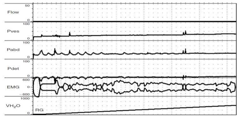
Proper history taking is necessary for appropriate evaluation of neurogenic bladder. Patients are asked whether he can sense the urge of micturition, able to evacuate the bladder voluntarily, experience any leakage in between, increased frequency, incomplete evacuation, unable to hold urine during urge or during coughing or abdominal pressure. Any history regarding fever, chills, sweating, blurred vision, raised blood pressure, redness of urine, bowel habit should be noted. Significant medical and surgical history specially any injury or surgical procedure of urinary tract should be asked. Occupational history of patient and history of functional achievementare also very important. Thorough physical examination specially abdomen, genitalia, perineum and per rectal examination is essential.Proper assessment of muscle power, spasticity (tightness), sensation, reflexes is required to plan proper rehabilitation protocol. Hand function, bed mobility, sitting & standing balance, wheelchair transfer are evaluated to determine how much dependency required for patients. Routine blood test is done for assessment of renal function and to rule out any infection &other complications. Urine analysis is essentialfor diagnosis and treatment of urinary tract infection and renal abnormalities. USG of KUB is required for assessment of pre and post-voidal residual urine (to assess ability of evacuation), bladder outlet obstruction and any structural abnormalities. Urodynamic study (Fig. 1) is very important diagnostic tool for objective assessment of detrusor contraction, intravesical pressure, urethral sphincter pressure (electromyography of sphinteric muscle), dyssynergy and uroflowmetry.In OAB, increased contraction of detrusor is seen during filling phase of urodynamic study (UDS). In UAB, lack or diminished contraction of detrusor is detected by voiding phase of urodynamic study. In Dyssynergia, both detrusor and sphincter contract simultaneously in both filling and voiding phase of UDS. For direct visualization of internal bladder wall, Cystoscopy is performed. It isindicated if there are hematuria, recurrent UTI, bladder stone, suspicion of malignancy or structural abnormality of bladder. CT/CT urogram may be done to evaluate for stone, tumour, microscopic hematuria etc.

How do you manage?

At first, a rehab goal is set by Neuro-Rehab physician (team leader) along with other team members like rehab nurse, physiotherapist, occupational therapist, prosthetist-orthotist, psychological& vocational counselor, social worker and patient with family members or care giver. Bladder rehabilitation protocol is customized according to goal and conditions of bladder. It consists of pharmacotherapy, behavioural therapy, supportive care, interventional procedure and surgical management.

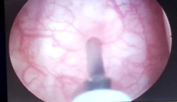
In overactive bladder, where urge incontinence is the main problem, time voiding practice is very helpful.Patientsare trained to void before they reach their full bladder capacity or maximum urge of micturition. They are also educated how to increase the time between voiding by 10 to 15 minutes every 2 to 5 days.A bladder diary should be maintained where amount of intake, urine output and amount of leakage are recorded. Supportive care with diaper, external condom catheter, intermittent catheterization are also suggested. Oral medications like Anti-cholinergic (darifenacin, solifenacin, oxybutynin, Tolterodine, TCA etc.) and Mirabegron are usually recommended. They all inhibit detrusor contraction as well as urge incontinence. Posterior Tibial nerve stimulation is a nonsurgical & non-painful procedure for OAB. Some studies suggested efficacy upto 80%. In this procedure, a slim needle electrode is inserted near the ankle in posterior tibial nerve and surface electrode is placed on foot. Electrodes are connected to a low voltage stimulator. Electrical impulse travels to tibial nerve and then to sacral nerve which controls bladder function. When bladder function is not optimum with these conservative management or there is serious adverse effect, various minimally invasive procedures are performed by Interventional Neuro Physiatrists independently or with collaboration of Urologist. Cystoscopy guided botulinum toxin injection to detrusor muscle (Fig. 2) is highly recommended. 100-300 units botulinum toxin type A is injected in 20-30 injection sites (10U/ml each) of bladder wall except trigone.Usually local or spinal anesthesia is sufficient for this interventional procedure. It takes 1 to 4 weeks to act and effectpersists up to 9 months.Significant improvement in detrusor pressure, compliance and bladder capacity are observed in these cases. Intravesical injections with lidocaine, oxybutynin, capsaicin are other options. If conservative and non-surgical procedures are ineffective, different urinary surgeries are performed by Urosurgeon.Surgical procedure like bladder augmentation (increase bladder capacity), detrusor myomectomy, urinary diversion are done in refractory cases.

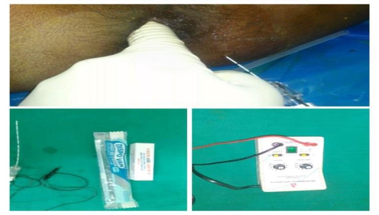
In DSD, urinary retention is the main problem. Anal stretching is effectivein these patients to decrease retention. Stretching of anal sphincter leads to relaxation of pelvic floor muscles which helps to empty bladder by Valsalva’s maneuver (increasing intraabdominal pressure). This technique is suggested if patient is able to transfer to toilet and perform Valsalva’s maneuver with absence of anal pain sensation. Timed voiding is ineffective hare. Various oral medications like alfa-1 blockers (terazosin, prazosin, tamsulosin, silodosin etc.), baclofen, diazepam, dantrolene sodium are effective. These drugs mainly relax smooth muscles of bladder neck, urethra and prostate. Some side effectslike postural hypotension,palpitation, headache, dizziness, nasal stuffiness etc. are commonly reported. When Non-Pharmacological and Pharmacological therapies are not sufficient or intolerant to drugs, interventional procedures are recommended.Botulinum toxin A injection (100 units) to external sphincter(4 sites) is very effective to relax external sphincteric muscles. It is a day care procedure, usually done under cystoscopy guidance with local or spinal anesthesia. It improves post-voidal residual urine (71% decrease), leak point pressure, maximal intravesical & urethral pressure within 3-4 weeks. Another technique is Pudendal nerve block (neurolysis) to inhibit abnormal contraction of external sphincteric muscles. Pudendal neurolysis is commonly performed with 5% aqueous phenol under guidance of small percutaneous direct current peripheral stimulator with 100mm Teflon coated needle (Fig. 3). It is a day care procedure, usually done with local anesthesia.Reduction of Post-voidal residual urine, leak point pressure, max detrusor pressure and improvement of bladder capacityare seen within 1 month


and maximum effect is seen at 3 months. When non-surgical treatments are inadequate, surgeries are the treatment of choice. Surgical procedures like transurethral sphincterotomy, urethral stent, bladder neck ablation etc. are planned.

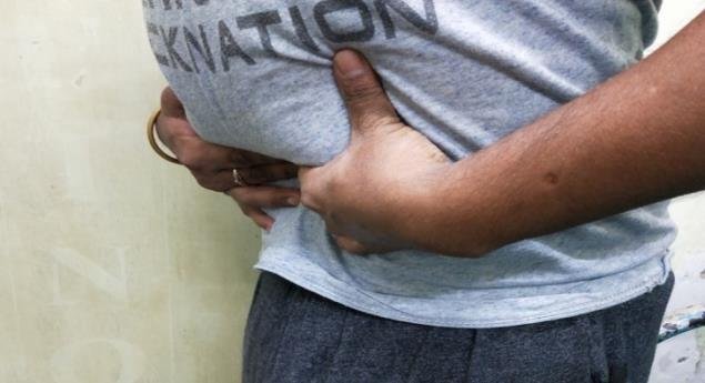
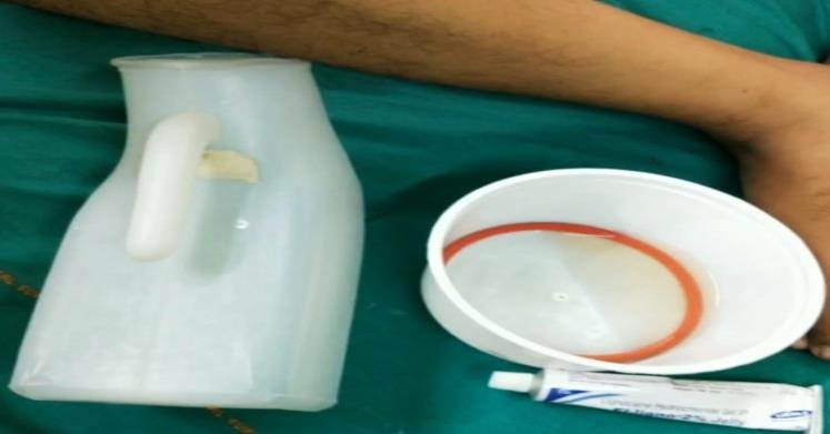
In case of underactive bladder, patient experiences urinary retention. Timed voiding has some effective roles along with increasing intravesical pressure by Valsalva’s or Crede’s maneuver. In Crede’s maneuver, closed fist is pushed down to the suprapubic area with enough force to express urine from the bladder (Fig.4).In Valsalva, it is bearing down with intra-abdominal pressure with enough force to push urine out of the bladder. These techniques are Best reserved for those who are unable to perform Intermittent catheterization, have decreased urethral sphincter activity such as                                                                                                                                                                           elderly women or SCI with LMN Lesion & sphincterotomy. Exacerbation of haemorrhoids, rectal prolapse,hernia etc. are the drawbacks in these techniques. In patients with weak uninhibited bladder contractions suprapubic bladder tapping may be used to trigger a contraction.. A successful simple method for management of failure to empty caused by the bladder in those with good hand function is clean intermittent catheterization (CIC) by self or by attendant if hand function of patient is poor. It is a simple non-painful technique, usually performed by rubber or Nelaton catheter without any gloves. Before CIC, proper preparation is required to avoid infection.Hands of patient (for self) or attendant (for assisted), catheter, one wide open container & urine pot are cleaned thoroughly with soap and water(Fig. 5). After applying lubricating gel over catheter thoroughly, catheter is inserted through urethral meatus by holding the penis forward for male or by seeing in mirror for female until urine begins to flow. After stopping urine flow, catheter is rotated and slowly removed. Then catheter is cleaned with water, dried in air and kept in dry wide-open container and reused. Usually, CIC is done five to six times in a day to avoid bladder overdistended along with fluid intake restricted to 1.5-2 liter per day. CIC is usually avoided in women with significant adductor spasticity, history of a urethral false passage, stricture and bladder neck obstruction, poor hand-eye coordination, poor cognition, poor motivation, inability or unwillingness to adhere to the catheterization time schedule or the fluid intake regimen, inability to catheterize themselves or a caregiver who is unable to perform catheterization. If there is lack of success to these methods or unable to CIC, indwelling urethral catheter is suggested and if there is urethral stricture, trauma or other contraindications, suprapubic catheterization is advised.Among oral medication, Bethanechol was recommended to increase detrusor contractility previously, but it is not used now a days due to adverse effect. Prostaglandin F2αis administered intravesically to increase detrusor pressures in SCI patients with supra-sacral lesions. More than 80% patients succeed to void with this procedure. Narcotic antagonists have also some role by stimulating sacral micturition reflex. To improve voiding by helping to trigger uninhibited contractions, electrodes may be placed on the bladder wall, pelvic nerves, sacral roots & conus medullaris. These are the surgical procedure and at present the only site being clinically used is sacral roots (on anterior roots).

Figure 1: Urodynamic study

Figure 2: Cystoscopy guided botulinum toxin injection to detrusor

Figure 3: Pudendal nerve chemoneurolysis with nerve stimulator

Figure 4: Crede’s maneuver

Figure 5: Preparation for clean intermittent catheterization (CIC)

Complex Regional Pain Syndrome and it’s management

BACKGROUND:

Complex Regional Pain Syndrome (CRPS) is one of the chronic severe and debilitating painful conditions attended in our PMR outdoor and indoor. First case of CRPS was reported in seventeenth century by French surgeon Ambroise when King Charles IX was suffering in persistent pain and contractures of arm after bloodletting. In American Civil War (1864), Silas Mitchell observed a chronic pain syndrome in soldiers who suffered traumatic injuries. He described this syndrome using the term ‘Causalgia’. At the beginning of the 20th century, Paul Sudeck identified the localized bone atrophy by x-rays which was

called ‘Sudeck’s atrophy’. In 1916 Rene Leriche suggested that Causalgia was caused by overactivity of sympathetic nervous system. He treated these patients with surgical sympathectomy. In 1950, John Bonica introduced the phrase ‘Reflex sympathetic dystrophy (RSD)’. International Association for the Study of Pain (IASP) differentiated RSD and causalgia in 1979.

WHAT IS CRPS?

C = Complex: Varied and dynamic clinical presentation

R = Regional: Non-dermatomal distribution of symptoms

P = Pain: Out of proportion to the inciting events

S = Syndrome: Constellation of symptoms and signs

It is a Disorder characterized by sympathetically maintained pain and related sensory abnormalities, abnormal blood flow, abnormalities in motor system and changes in both superficial and deep structures with trophic changes. CRPS is of two types:

CRPS type I (also known as RSD) often involves only one limb with pain, minor edema, after a period of immobilization.

CRPS type II (also called causalgia) is seen in presence of a known injury to a nerve.

WHO ARE SUFFERING MOST?

CRPS is seen most commonly in 45 -55 years age group, but patient with any age can suffer and more frequently affects women (70 – 80%). 60% of CRPS cases involve in the upper extremities. Incidence of CRPS ranges from 5.46 to 26.2 per 1,00,000 persons year. Prevalence subsequent to trauma ranges from

0.03 to 37% based on retrospective studies. Risk factors are smoking, post-menopausal female, adult age, immobilization, spasticity.

WHAT ARE THE CAUSES OF CRPS?

Causes of CRPS are not well understood. But it occurs following various clinical conditions, like Fracture (16-46%), Strain or sprain (10%–29%), Post surgery (3%–24%), Contusion or crush injury (8%–18%), Stroke (10-48%), Spinal cord disorders (6%), Repetitive motion disorder (carpal tunnel syndrome), Myocardial infarction, Burn, Infection (leprosy), Radiation therapy etc. There is no correlation between

the severity of trauma and the degree of CRPS symptoms. No psychological factor predisposing for CRPS has been identified. However, 80% of CRPS patients had ‘stressful life events’ close to the time of

diagnosis.

PATHOPHYSIOLOGY:

Exact mechanism of CRPS is not clear. However, three hypotheses are mainly proposed: neurogenic inflammation, vasomotor dysfunction and maladaptive neuroplasticity. Neurogenic inflammation causes swelling in central nervous system and nociceptive sensitization (extreme sensitivity or allodynia). Blood flow disorder due to alteration of sympathetic nervous system causes swelling and discolouration of

limb. Brain adapts constant pain signals. CRPS is result of an inappropriate response to tissue injury.

CLINICAL PRESENTATION:

Characteristic triad of symptoms are autonomic, sensory and motor disturbances. Pain is the hallmark of the disease. Allodynia, hyperesthesia & hyperalgesia are usually seen. Distal limbedema and skin

temperature are raised in 80% cases. Skin color is initially red, but becomes pale in chronic cases.

Sweating, nail and hair changes (increased growth), disuse atrophy, weakness, distal tremor and cramp are mostly noticed. Patients present also present with mood disturbances (anxiety, depression) and sleepdisturbances. Contralateral limb even can progress to all 4 limbs

LANKFORD AND EVANS STAGES

Acute stage (0-3 months): In this stage, patients present with severe burning pain, pitting oedema, warmth, reddish skin, hyperesthesia, increased hair and nail growth, joint stiffness.

Subacute or dystrophic stage (3-12 months): In this stage, pain worsens.Brawny oedema, pallor, ridged nail, cyanosis, stiffness are also seen.

Chronic or atrophic stage (> 12 months): Pain reduces in this stage.Glossy skin, joint contractures, atrophy are presented.

DIAGNOSIS:

CRPS is primarily diagnosed by clinical sign and symptoms. For clinical diagnosis,Budapest criteria(2003) were more sensitive and specific. Following fours criteria (A-D) must be met for diagnosis:

  1. Continuing pain which is disproportionate to any inciting event
  2. At least one symptom in three or more categories
    • Sensory – Hyperesthesia, allodynia
    • Vasomotor – temperature asymmetry, skin colour changes/asymmetry
    • Sudomotor/oedema –Oedema, sweating changes/asymmetry
    • Motor/trophic – Decreased ROM, motor dysfunction(weakness, tremor, dystonia), trophic changes (hair, nail, skin).
  3. At least one sign in two or more categories
    • Sensory – hyperalgesia (pinprick), allodynia (lighttouch/temp/pressure)
    • Vasomotor – temperature asymmetry (> 1 °C), skin colour changes/asymmetry


  • Sudomotor/oedema –oedema, sweating changes/asymmetry
  • Motor/trophic – decreased ROM, motor dysfunction (weakness, tremor, dystonia), trophic changes (hair, nail, skin)
  1. No other diagnosis can better explain the signs and symptoms

DIFFERENTIAL DIAGNOSIS:

There are other diseases with similar clinical presentations. Cellulitis, Lymphedema, Stress fracture, Acute synovitis, Septic arthritis, Tenosynovitis, Thrombophlebitis, DVT, PVD, Arterial insufficiency, Dupuytren’s disease, Post herpetic neuralgia, Vasculitis should be differentiated from CRPS.

INVESTIGATIONS:

  • Blood tests: Routine blood and inflammatory markers are tested to rule out underlying infection or inflammatory arthritis.
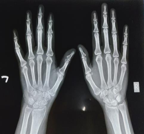
  • X- Ray of both hands/feet: Although there is no change in X-ray at acute stage, subchondral

osteopenia (fig.2) is seen in subacute stage and extreme osteopenia/osteoporosis is noted in chronic stage.

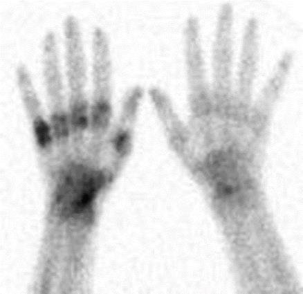
  • Three-phase bone scintigraphy (fig.4): It is highly sensitive and specific for Significant uptake is seen in the metacarpal bones specially in the subacute stage.
  • Sympathetic ganglion diagnostic block: This is a diagnostic block by local anesthetic agents to

confirm whether it is sympathetic mediated pain or not. This is performed usually under fluoroscopy or USG guidance as vital structures are present nearby.

  • Regional intravenous diagnostic block & Phentolamine infusion test (alpha-1 receptor antagonist) are other diagnostic procedures.
  • To exclude other diagnosis, MRI (may show marrow oedema but not specific for CRPS), EMG- NCS, Doppler flow meter, Vascular scintigraphy, Vital capillaroscopy are performed.

TREATMENT:

Early recognition is the key of management. Treatment of CRPS requires an individualized

comprehensive rehabilitation protocol on holistic approach to achieve complete recovery and prevent from progressing to irreversible stage of CRPS (Pachydermia). Comprehensive rehabilitation protocol consists of pharmacological therapy (medication), Nonpharmacological therapy (Physiotherapy, occupational therapy etc.) & interventional procedure. Surgery is the treatment of choice when

conservative management fails.

Pharmacological therapy: Following medicines are initially prescribed for management of pain, inflammation or swelling and demineralization:-

  • Non-steroidal anti-inflammatory drugs (NSAIDs): first line treatment in early stage, although their definitive efficacy is not proven.
  • Bisphosphonates: Oral and intravenous both bisphosphonates (Risedronate, palmidronate ) are primarily used for bone mineralization. They significantly relieve spontaneous pain and improve functional status in early stage.
  • Oral corticosteroids: Short course of steroid may be indicated in acute stage for their anti-inflammatory properties.
  • Tricyclic antidepressant (Amitriptyline, Nortriptyline), Gabapentin and Pregabalin may be They act as adjuvants by inhibiting pain pathways & neuronal plasticity.
  • Opioids like N-methyl-D-aspartate (NMDA) receptor antagonists (Ketamine, Methadone) are effective in CRPS-I due to phenomenon of central sensitization.
  • Vitamin C and N-acetylcysteine have some preventive role for CRPS in wrist fracture
  • Dimethyl sulfoxide 50% cream reduce pain & inflammation in acute

Non pharmacological therapy: Apart from medications, role of non-pharmacological therapy is utmost important. Therapeutic exercises like Joint Range of motion exercises, stretching & strengthening

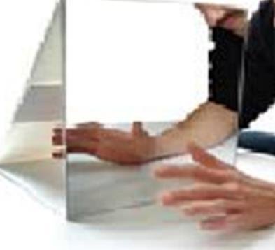
exercises are advised primarily to improve limb mobility. TENS, Paraffin bath & Hyperbaric oxygen therapy have significant role in management of pain, swelling & stiffness. rTMS is suggested in some studies for neuromodulation. Desensitization technique, Hand function training and gait training are important for functional improvement. There are some positive evidences of mirror therapy (fig.5), Qigong, acupuncture for treatment of CRPS.

Interventional procedures: When noninvasive methods are not responsive, interventional procedures are planned. Following regional procedures are strongly recommended by various literatures:

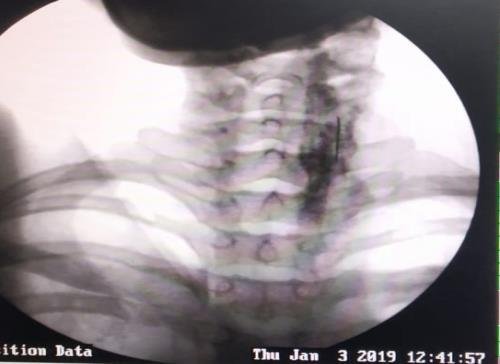
  • Stellate ganglion block (fig.6): For upper limb CRPS, stellate ganglion block with local anesthetic

agent and corticosteroid is the treatment of choice. It is a day care procedure usually done under USG or fluoroscopy guidance. Stellate ganglion is located anterior to transverse process of C7 and neck of first rib. Patient is positioned supine with a thin pillow under neck. Head rotated slightly to the opposite side. On fluoroscopic technique, needle is directed to the junction of vertebral body

and transverse process of C6 or C7 between trachea and carotid artery. After bony contact, needle is slightly withdrawn to rest outside of longus coli muscle. Contrast agent is injected to confirm

appropriate needle position and to rule out intravascular or neuraxial injection. After confirmation by both antero-posterior & lateral view, mixture of local anesthetic agent and corticosteroid is

injected. Onset of Horner syndrome (Ptosis, miosis, anhidrosis) with increased temperature (>3F) of affected limb is usually observed in successful block. On USG guided technique, transducer is placed over C6 level. After retracting carotid artery laterally, needle is inserted paratracheally using an in-

plane approach. Following a negative aspiration test for blood or CSF, local anesthetic agent and corticosteroid are injected to subfascial plane of prevertebral fascia.

  • Lumbar sympathetic block (fig.7): Fluoroscopy or USG guided lumbar sympathetic ganglion block is recommended for lower limb CRPS. On fluoroscopic technique, patient is placed on prone position with a pillow under lower abdomen. After dressing & draping, needle is inserted towards antero-lateral side of L2 or L3 vertebral body. After confirmation with contrast agent, mixture of local

anesthetic agent and corticosteroid is injected. On USG guided approach, patient is positioned on lateral decubitus with affected limb upwards. Needle is inserted below the lower pole of kidney towards antero-lateral aspect of L2 or L3 vertebral body. After repeated negative aspiration, local anesthetic & corticosteroid are injected on real time visualization. Patient is monitored for 2 hrs and may be discharged after 4 hrs.

  • Radiofrequency ablation (RFA) of sympathetic ganglion: Both conventional and pulsed RFA are indicated for long term effect and in cases where corticosteroid is contraindicated.
  • Bier block: Some studies suggest intravenous injection of local anesthesia and corticosteroid for reduction of pain in CRPS-1.

Surgical procedures:

Surgery is the treatment of choice in patients refractory to conservative therapy and interventional procedures. Spinal cord stimulation, percutaneous cervicothoracic or lumbar sympathectomy and intrathecal pump of morphine are the effective surgical procedures for CRPS.

PROGNOSIS:

Prognosis of CRPS is highly variable. Better outcome is seen in younger persons, children and teenagers. For older people, good outcome may be noticed but some persons experience severe pain and disability despite treatment. Evidences suggest early treatment, particularly rehabilitation, is helpful in limiting

disability.

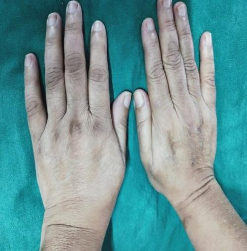
Figure 1: Left upper limb CRPS following supracondylar fracture

Figure 2: X-ray of both hand (AP view) in left upper limb CRPS

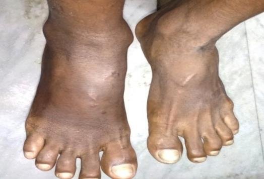
Figure 3: Right lower limb CRPS

Figure 4: Three-phase bone scintigraphy in left upper limb CRPS

Figure 5: Mirror therapy

Figure 6: Stellate ganglion block under fluoroscopy guidance

Figure 7: Fluoroscopic guided lumbar sympathetic ganglion block

Heel Pain Causes, Symptoms, Treatment, and When to See a Doctor

Heel Pain: Don’t Ignore Your First Steps

Heel pain is one of the most common foot complaints affecting people of all ages. It can make simple activities like getting out of bed, walking to work, climbing stairs, or standing for long periods difficult and painful. Although many people ignore heel pain initially, early diagnosis and treatment can prevent the condition from becoming chronic.

This article explains the common causes of heel pain, symptoms to watch for, available treatment options, and practical ways to prevent it.

What Is Heel Pain?

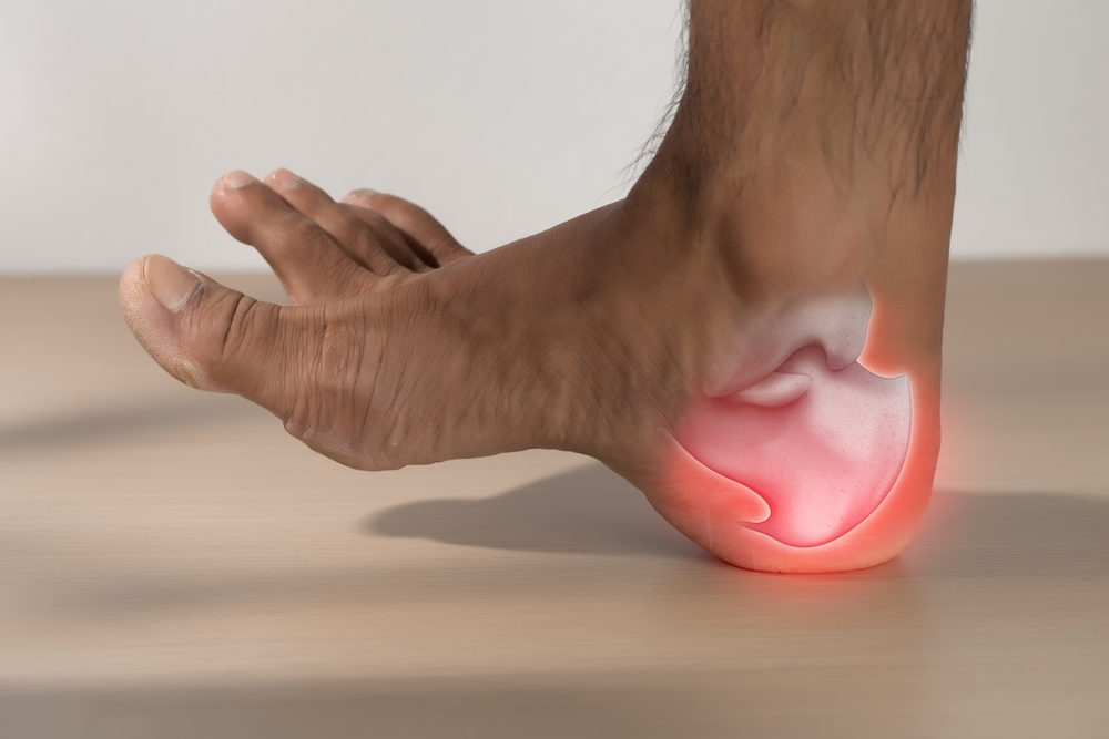
Heel pain refers to discomfort occurring underneath, behind, or around the heel bone (calcaneus). The pain may develop gradually due to repeated stress or appear suddenly after an injury.

Most cases respond well to conservative treatment, but persistent pain should be evaluated by a healthcare professional.

Common Causes of Heel Pain

  1. Plantar Fasciitis

The most common cause of heel pain is plantar fasciitis. The plantar fascia is a thick band of tissue that supports the arch of the foot. Repeated strain causes small tears and inflammation.

Typical symptoms include:

  • Sharp pain during the first few steps in the morning
  • Pain after prolonged sitting
  • Improvement after walking for a few minutes
  • Pain returning after long periods of standing
  1. Heel Spur

A heel spur is a calcium deposit that forms on the heel bone. Many heel spurs do not cause symptoms, but they may coexist with plantar fasciitis and contribute to discomfort.

  1. Achilles Tendinitis

Inflammation of the Achilles tendon causes pain behind the heel. It is common in runners, athletes, and people who suddenly increase their physical activity.

Symptoms include:

  • Pain behind the heel
  • Swelling
  • Morning stiffness
  • Pain while climbing stairs
  1. Heel Pad Syndrome

The natural fat pad beneath the heel becomes thinner or bruised, leading to pain when walking on hard surfaces.

  1. Stress Fracture

Repeated impact activities such as running or jumping may cause tiny cracks in the heel bone.

  1. Nerve Entrapment

Compression or irritation of nerves around the ankle can produce burning pain, tingling, or numbness in the heel.

Who Is at Higher Risk?

Heel pain is more common in:

  • People who stand for long hours
  • Teachers
  • Factory workers
  • Healthcare professionals
  • Athletes
  • Runners
  • People who are overweight
  • Individuals with flat feet or high arches
  • Older adults

Symptoms of Heel Pain

Depending on the cause, symptoms may include:

  • Sharp pain under the heel
  • Pain with the first steps in the morning
  • Pain after prolonged standing
  • Burning sensation
  • Swelling
  • Tenderness
  • Difficulty walking
  • Pain after exercise

Warning Signs That Need Medical Attention

Consult a doctor if you experience:

  • Heel pain lasting more than 2–3 weeks
  • Severe swelling
  • Inability to bear weight
  • Fever associated with heel pain
  • Redness and warmth around the heel
  • Numbness or weakness
  • Pain after a significant injury

Early diagnosis helps identify the underlying cause and allows timely treatment.

How Is Heel Pain Diagnosed?

Your doctor may perform:

  • Detailed medical history
  • Physical examination
  • Assessment of walking pattern
  • Foot alignment evaluation
  • X-ray (if fracture or heel spur is suspected)
  • Ultrasound (to evaluate soft tissue structures)
  • MRI (for selected cases where soft tissue injury or stress fracture is suspected)

Treatment Options

Treatment depends on the underlying cause.

Rest and Activity Modification

Reducing activities that worsen the pain allows healing.

Ice Therapy

Applying an ice pack for 15–20 minutes several times a day can reduce pain and inflammation.

Stretching Exercises

Regular stretching of the calf muscles and plantar fascia helps relieve symptoms and improve flexibility.

Proper Footwear

Supportive shoes with good arch support and cushioning reduce stress on the heel.

Orthotic Insoles

Custom or prefabricated insoles may help distribute pressure more evenly across the foot.

Medications

Your doctor may prescribe medications to relieve pain and inflammation when appropriate.

Physiotherapy

A supervised rehabilitation program can improve flexibility, strengthen supporting muscles, correct gait abnormalities, and reduce the likelihood of recurrence.

Image-Guided Interventions

In carefully selected patients with persistent heel pain that has not responded to conservative treatment, ultrasound-guided injections or other image-guided pain procedures may be considered. These should only be performed after a thorough clinical evaluation and discussion of potential benefits and risks.

Surgery

Surgery is rarely required and is usually reserved for patients whose symptoms persist despite several months of appropriate non-surgical treatment.

Can Heel Pain Be Prevented?

Yes. Simple preventive measures include:

  • Maintain a healthy body weight.
  • Wear supportive footwear.
  • Replace worn-out shoes regularly.
  • Stretch your calf muscles daily.
  • Avoid prolonged standing whenever possible.
  • Increase exercise intensity gradually.
  • Warm up before sports.
  • Avoid walking barefoot on hard floors if it aggravates your symptoms.

Frequently Asked Questions (FAQ)

Is heel pain always caused by a heel spur?

No. Plantar fasciitis is a much more common cause of heel pain. Many people have heel spurs without experiencing any symptoms.

Can heel pain go away on its own?

Some mild cases improve with rest and stretching. Persistent or worsening pain should be evaluated by a doctor.

Is walking good for heel pain?

Gentle walking may be acceptable for some people, but excessive walking during painful periods can worsen symptoms. Your doctor or physiotherapist can advise you based on the cause.

How long does heel pain take to improve?

Recovery varies depending on the diagnosis. Many patients improve over weeks to a few months with appropriate treatment and rehabilitation.

When Should You Visit a Pain Specialist?

If heel pain continues despite rest, home exercises, or over-the-counter measures, or if it interferes with your daily activities, seeking medical evaluation is important. A pain specialist can identify the underlying cause, recommend evidence-based treatment, and develop a personalized rehabilitation plan.

Conclusion

Heel pain should never be ignored, especially when it becomes persistent or limits your daily activities. Most causes are treatable with early diagnosis, appropriate rehabilitation, and lifestyle modifications. Seeking timely medical advice can help relieve pain, restore mobility, and prevent long-term complications.

If you are experiencing persistent heel pain, consult a qualified healthcare professional for a comprehensive assessment and individualized treatment plan.