Rehabilitation Of Neurogenic Urinary Bladder In Spinal Cord Injury
- Home
- Rehabilitation Of Neurogenic Urinary Bladder In Spinal Cord Injury
A spinal cord injury (SCI) may interrupt the neural connection between brain and spinal nerves controlling urinary bladder function. This can lead to dysfunctional voiding process which is also known as Neurogenic bladder. Neurogenic bladder may cause loss of sensation as well as urge of micturition, inability to empty bladder, increase urinary frequency, leakage etc. These conditions can have a negative effect on quality of life as well as complications like urinary tract infection, kidney stone, Autonomic Dysreflexia. A comprehensive rehabilitation protocol can help patients to regain some control over bladder function and improve quality of life.
Lower urinary tract is supplied by three sets of peripheral nerves: parasympathetic, sympathetic and pudendal nerve. Pelvic nerves which originate from S2-S4 sacral spinal roots are parasympathetic nerves. They cause contraction of bladder wall muscle (detrusor) and relaxes internal urethral sphincter. So stimulation of these nerves lead to emptying of bladder or voiding. Hypogastric nerves originating from T10-L2 spinal roots are the main sympathetic nerves. They cause relaxation of detrusor and contraction of internal sphincter. So, there is storage of urine due to stimulation of these sympathetic nerves. Pudendal nerves arising from S2-S4 are the main somatic nerves and innervate external urethral sphincter and pelvic floor muscles. Pudendal nerves are under voluntary control, whereas sympathetic and parasympathetic nerves act automatically.
Sacral micturition center is located at S2-S4 segments of spinal cord through which bladder can contract independently. Brain sends signal to sacral micturition centre telling the bladder when and where not to contract. Normal micturition is a spinal reflex modulated by central nervous system (brain and spinal cord) which coordinates bladder and urethra.
Two types of bladder disorders are seen in SCI : immediately after injury (in spinal shock stage), and abnormalities (long term) after spinal shock stage.
During spinal shock stage, signals from the brain cannot transmit to the parts below the spinal cord injury and bladder cannot be emptied. It usually lasts for up to a few days, but for the bladder it can last several months or longer.
After spinal shock, bladder dysfunction depends on level of spinal cord injury. In case of SCI above sacral micturition center, signals (when and where not to contract the bladder) normally come down from brain& brain stem are blocked. Therefore, detrusor begins to contract and possibly cause urinate without control (urinary incontinence). This is called as Overactive Bladder (OAB). Internal sphincter might tighten or relax on its own, starting and stopping urine stream without control. Bladder might try to squeeze but the sphincter might tighten at the same time, making unable to urinate and causing a high-pressure buildup in the bladder (urinary retention). This is known as Detrusor Sphincter Dyssynergia (DSD).
If the SCI damaged the spinal cord at or near the base of spine, the sacral micturition center might be damaged. So, signals cannot be sent to the bladder to tell the bladder to squeeze. This condition is known as Underactive Bladder (UAB). If the damage is below sacral micturition center, even though signals are sent towards the bladder, the nerves to the bladder are damaged so the signals do not
reach the bladder causing UAB. In this condition, bladder may become over distended. If there is weak urinary sphincter or bladder pressure overcomes sphincteric pressure, urine will overflow from bladder without control. This is known as overflow incontinence.
One of common complication of these patients is urinary tract infection (UTI). It may often present as asymptomatic or symptoms like fever, chills, back pain, lower abdominal pain, pain during micturition, frequency and testicular swelling. Renal stone is also commonly seen in these patients mostly due to infection and altered urinary pH. Long term indwelling catheter may increase incidence of bladder cancer. Noxious stimuli like UTI, 0verdistension of bladder or catheter blockage may trigger to Autonomic Dysreflexia (AD) in SCI at T6 or above level. AD is a potentially dangerous syndrome causing sudden and severe rise of blood pressure (>20 mmHg over baseline SBP). It usually presents as mild symptoms like headache, blurred vision, sweating, goosebumps, nasal stuffiness to life threatening complications like seizure, stroke, myocardial infraction and retinal detachment. Immediate reversal of noxious stimulus and rapid control of blood pressure are required for management of this fatal medical emergency. Social isolation due to urinary leakage and wetting clothes may lead to depression.
Reintegration of patient into the community by providing a socially acceptable way of voiding is the main goal. Bladder should not be evacuated more than every 3 to 4 hours. It should remain socially continent. Sleep should not be interfered with incontinence. Prevention and early detection of upper and lower urinary tract complications are very important for rehabilitation of neurogenic bladder.
Proper history taking is necessary for appropriate evaluation of neurogenic bladder. Patients are asked whether he can sense the urge of micturition, able to evacuate the bladder voluntarily, experience any leakage in between, increased frequency, incomplete evacuation, unable to hold urine during urge or during coughing or abdominal pressure. Any history regarding fever, chills, sweating, blurred vision, raised blood pressure, redness of urine, bowel habit should be noted. Significant medical and surgical history specially any injury or surgical procedure of urinary tract should be asked. Occupational history of patient and history of functional achievementare also very important. Thorough physical examination specially abdomen, genitalia, perineum and per rectal examination is essential.Proper assessment of muscle power, spasticity (tightness), sensation, reflexes is required to plan proper rehabilitation protocol. Hand function, bed mobility, sitting & standing balance, wheelchair transfer are evaluated to determine how much dependency required for patients. Routine blood test is done for assessment of renal function and to rule out any infection &other complications. Urine analysis is essentialfor diagnosis and treatment of urinary tract infection and renal abnormalities. USG of KUB is required for assessment of pre and post-voidal residual urine (to assess ability of evacuation), bladder outlet obstruction and any structural abnormalities. Urodynamic study (Fig. 1) is very important diagnostic tool for objective assessment of detrusor contraction, intravesical pressure, urethral sphincter pressure (electromyography of sphinteric muscle), dyssynergy and uroflowmetry.In OAB, increased contraction of detrusor is seen during filling phase of urodynamic study (UDS). In UAB, lack or diminished contraction of detrusor is detected by voiding phase of urodynamic study. In Dyssynergia, both detrusor and sphincter contract simultaneously in both filling and voiding phase of UDS. For direct visualization of internal bladder wall, Cystoscopy is performed. It isindicated if there are hematuria, recurrent UTI, bladder stone, suspicion of malignancy or structural abnormality of bladder. CT/CT urogram may be done to evaluate for stone, tumour, microscopic hematuria etc.
At first, a rehab goal is set by Neuro-Rehab physician (team leader) along with other team members like rehab nurse, physiotherapist, occupational therapist, prosthetist-orthotist, psychological& vocational counselor, social worker and patient with family members or care giver. Bladder rehabilitation protocol is customized according to goal and conditions of bladder. It consists of pharmacotherapy, behavioural therapy, supportive care, interventional procedure and surgical management.
In overactive bladder, where urge incontinence is the main problem, time voiding practice is very helpful.Patientsare trained to void before they reach their full bladder capacity or maximum urge of micturition. They are also educated how to increase the time between voiding by 10 to 15 minutes every 2 to 5 days.A bladder diary should be maintained where amount of intake, urine output and amount of leakage are recorded. Supportive care with diaper, external condom catheter, intermittent catheterization are also suggested. Oral medications like Anti-cholinergic (darifenacin, solifenacin, oxybutynin, Tolterodine, TCA etc.) and Mirabegron are usually recommended. They all inhibit detrusor contraction as well as urge incontinence. Posterior Tibial nerve stimulation is a nonsurgical & non-painful procedure for OAB. Some studies suggested efficacy upto 80%. In this procedure, a slim needle electrode is inserted near the ankle in posterior tibial nerve and surface electrode is placed on foot. Electrodes are connected to a low voltage stimulator. Electrical impulse travels to tibial nerve and then to sacral nerve which controls bladder function. When bladder function is not optimum with these conservative management or there is serious adverse effect, various minimally invasive procedures are performed by Interventional Neuro Physiatrists independently or with collaboration of Urologist. Cystoscopy guided botulinum toxin injection to detrusor muscle (Fig. 2) is highly recommended. 100-300 units botulinum toxin type A is injected in 20-30 injection sites (10U/ml each) of bladder wall except trigone.Usually local or spinal anesthesia is sufficient for this interventional procedure. It takes 1 to 4 weeks to act and effectpersists up to 9 months.Significant improvement in detrusor pressure, compliance and bladder capacity are observed in these cases. Intravesical injections with lidocaine, oxybutynin, capsaicin are other options. If conservative and non-surgical procedures are ineffective, different urinary surgeries are performed by Urosurgeon.Surgical procedure like bladder augmentation (increase bladder capacity), detrusor myomectomy, urinary diversion are done in refractory cases.
In DSD, urinary retention is the main problem. Anal stretching is effectivein these patients to decrease retention. Stretching of anal sphincter leads to relaxation of pelvic floor muscles which helps to empty bladder by Valsalva’s maneuver (increasing intraabdominal pressure). This technique is suggested if patient is able to transfer to toilet and perform Valsalva’s maneuver with absence of anal pain sensation. Timed voiding is ineffective hare. Various oral medications like alfa-1 blockers (terazosin, prazosin, tamsulosin, silodosin etc.), baclofen, diazepam, dantrolene sodium are effective. These drugs mainly relax smooth muscles of bladder neck, urethra and prostate. Some side effectslike postural hypotension,palpitation, headache, dizziness, nasal stuffiness etc. are commonly reported. When Non-Pharmacological and Pharmacological therapies are not sufficient or intolerant to drugs, interventional procedures are recommended.Botulinum toxin A injection (100 units) to external sphincter(4 sites) is very effective to relax external sphincteric muscles. It is a day care procedure, usually done under cystoscopy guidance with local or spinal anesthesia. It improves post-voidal residual urine (71% decrease), leak point pressure, maximal intravesical & urethral pressure within 3-4 weeks. Another technique is Pudendal nerve block (neurolysis) to inhibit abnormal contraction of external sphincteric muscles. Pudendal neurolysis is commonly performed with 5% aqueous phenol under guidance of small percutaneous direct current peripheral stimulator with 100mm Teflon coated needle (Fig. 3). It is a day care procedure, usually done with local anesthesia.Reduction of Post-voidal residual urine, leak point pressure, max detrusor pressure and improvement of bladder capacityare seen within 1 month
and maximum effect is seen at 3 months. When non-surgical treatments are inadequate, surgeries are the treatment of choice. Surgical procedures like transurethral sphincterotomy, urethral stent, bladder neck ablation etc. are planned.
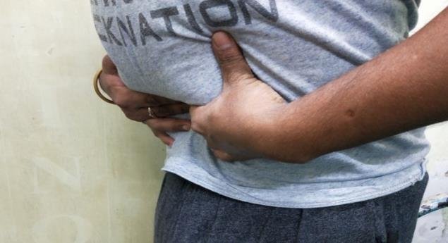
In case of underactive bladder, patient experiences urinary retention. Timed voiding has some effective roles along with increasing intravesical pressure by Valsalva’s or Crede’s maneuver. In Crede’s maneuver, closed fist is pushed down to the suprapubic area with enough force to express urine from the bladder (Fig.4).In Valsalva, it is bearing down with intra-abdominal pressure with enough force to push urine out of the bladder. These techniques are Best reserved for those who are unable to perform Intermittent catheterization, have decreased urethral sphincter activity such as elderly women or SCI with LMN Lesion & sphincterotomy. Exacerbation of haemorrhoids, rectal prolapse,hernia etc. are the drawbacks in these techniques. In patients with weak uninhibited bladder contractions suprapubic bladder tapping may be used to trigger a contraction.. A successful simple method for management of failure to empty caused by the bladder in those with good hand function is clean intermittent catheterization (CIC) by self or by attendant if hand function of patient is poor. It is a simple non-painful technique, usually performed by rubber or Nelaton catheter without any gloves. Before CIC, proper preparation is required to avoid infection.Hands of patient (for self) or attendant (for assisted), catheter, one wide open container & urine pot are cleaned thoroughly with soap and water(Fig. 5). After applying lubricating gel over catheter thoroughly, catheter is inserted through urethral meatus by holding the penis forward for male or by seeing in mirror for female until urine begins to flow. After stopping urine flow, catheter is rotated and slowly removed. Then catheter is cleaned with water, dried in air and kept in dry wide-open container and reused. Usually, CIC is done five to six times in a day to avoid bladder overdistended along with fluid intake restricted to 1.5-2 liter per day. CIC is usually avoided in women with significant adductor spasticity, history of a urethral false passage, stricture and bladder neck obstruction, poor hand-eye coordination, poor cognition, poor motivation, inability or unwillingness to adhere to the catheterization time schedule or the fluid intake regimen, inability to catheterize themselves or a caregiver who is unable to perform catheterization. If there is lack of success to these methods or unable to CIC, indwelling urethral catheter is suggested and if there is urethral stricture, trauma or other contraindications, suprapubic catheterization is advised.Among oral medication, Bethanechol was recommended to increase detrusor contractility previously, but it is not used now a days due to adverse effect. Prostaglandin F2αis administered intravesically to increase detrusor pressures in SCI patients with supra-sacral lesions. More than 80% patients succeed to void with this procedure. Narcotic antagonists have also some role by stimulating sacral micturition reflex. To improve voiding by helping to trigger uninhibited contractions, electrodes may be placed on the bladder wall, pelvic nerves, sacral roots & conus medullaris. These are the surgical procedure and at present the only site being clinically used is sacral roots (on anterior roots).
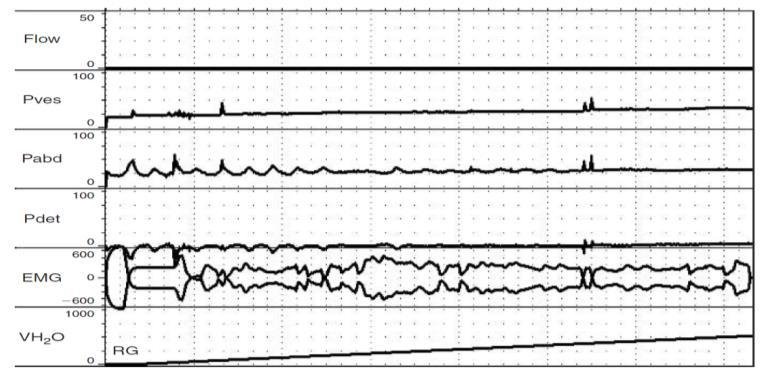
Figure 1: Urodynamic study
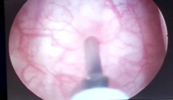
Figure 2: Cystoscopy guided botulinum toxin injection to detrusor
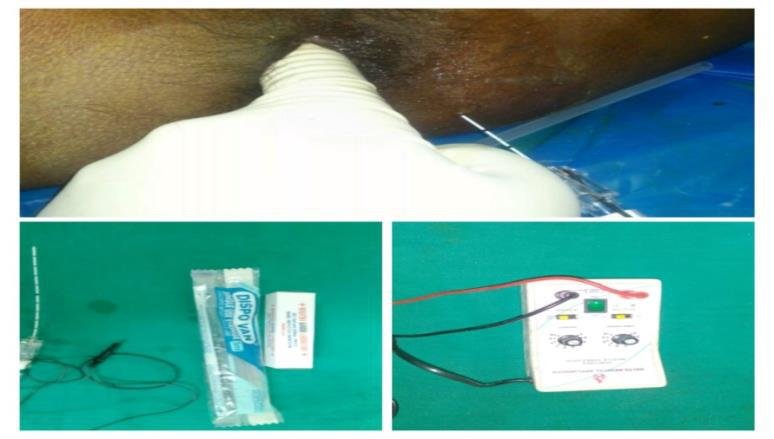
Figure 3: Pudendal nerve chemoneurolysis with nerve stimulator
Figure 4: Crede’s maneuver
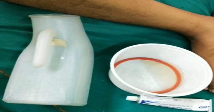
Figure 5: Preparation for clean intermittent catheterization (CIC)