Complex Regional Pain Syndrome and it’s management
- Home
- Complex Regional Pain Syndrome and it’s management
Complex Regional Pain Syndrome (CRPS) is one of the chronic severe and debilitating painful conditions attended in our PMR outdoor and indoor. First case of CRPS was reported in seventeenth century by French surgeon Ambroise when King Charles IX was suffering in persistent pain and contractures of arm after bloodletting. In American Civil War (1864), Silas Mitchell observed a chronic pain syndrome in soldiers who suffered traumatic injuries. He described this syndrome using the term ‘Causalgia’. At the beginning of the 20th century, Paul Sudeck identified the localized bone atrophy by x-rays which was
called ‘Sudeck’s atrophy’. In 1916 Rene Leriche suggested that Causalgia was caused by overactivity of sympathetic nervous system. He treated these patients with surgical sympathectomy. In 1950, John Bonica introduced the phrase ‘Reflex sympathetic dystrophy (RSD)’. International Association for the Study of Pain (IASP) differentiated RSD and causalgia in 1979.
C = Complex: Varied and dynamic clinical presentation
R = Regional: Non-dermatomal distribution of symptoms
P = Pain: Out of proportion to the inciting events
S = Syndrome: Constellation of symptoms and signs
It is a Disorder characterized by sympathetically maintained pain and related sensory abnormalities, abnormal blood flow, abnormalities in motor system and changes in both superficial and deep structures with trophic changes. CRPS is of two types:
CRPS type I (also known as RSD) often involves only one limb with pain, minor edema, after a period of immobilization.
CRPS type II (also called causalgia) is seen in presence of a known injury to a nerve.
CRPS is seen most commonly in 45 -55 years age group, but patient with any age can suffer and more frequently affects women (70 – 80%). 60% of CRPS cases involve in the upper extremities. Incidence of CRPS ranges from 5.46 to 26.2 per 1,00,000 persons year. Prevalence subsequent to trauma ranges from
0.03 to 37% based on retrospective studies. Risk factors are smoking, post-menopausal female, adult age, immobilization, spasticity.
Causes of CRPS are not well understood. But it occurs following various clinical conditions, like Fracture (16-46%), Strain or sprain (10%–29%), Post surgery (3%–24%), Contusion or crush injury (8%–18%), Stroke (10-48%), Spinal cord disorders (6%), Repetitive motion disorder (carpal tunnel syndrome), Myocardial infarction, Burn, Infection (leprosy), Radiation therapy etc. There is no correlation between
the severity of trauma and the degree of CRPS symptoms. No psychological factor predisposing for CRPS has been identified. However, 80% of CRPS patients had ‘stressful life events’ close to the time of
diagnosis.
Exact mechanism of CRPS is not clear. However, three hypotheses are mainly proposed: neurogenic inflammation, vasomotor dysfunction and maladaptive neuroplasticity. Neurogenic inflammation causes swelling in central nervous system and nociceptive sensitization (extreme sensitivity or allodynia). Blood flow disorder due to alteration of sympathetic nervous system causes swelling and discolouration of
limb. Brain adapts constant pain signals. CRPS is result of an inappropriate response to tissue injury.
Characteristic triad of symptoms are autonomic, sensory and motor disturbances. Pain is the hallmark of the disease. Allodynia, hyperesthesia & hyperalgesia are usually seen. Distal limbedema and skin
temperature are raised in 80% cases. Skin color is initially red, but becomes pale in chronic cases.
Sweating, nail and hair changes (increased growth), disuse atrophy, weakness, distal tremor and cramp are mostly noticed. Patients present also present with mood disturbances (anxiety, depression) and sleepdisturbances. Contralateral limb even can progress to all 4 limbs
Acute stage (0-3 months): In this stage, patients present with severe burning pain, pitting oedema, warmth, reddish skin, hyperesthesia, increased hair and nail growth, joint stiffness.
Subacute or dystrophic stage (3-12 months): In this stage, pain worsens.Brawny oedema, pallor, ridged nail, cyanosis, stiffness are also seen.
Chronic or atrophic stage (> 12 months): Pain reduces in this stage.Glossy skin, joint contractures, atrophy are presented.
CRPS is primarily diagnosed by clinical sign and symptoms. For clinical diagnosis,Budapest criteria(2003) were more sensitive and specific. Following fours criteria (A-D) must be met for diagnosis:
There are other diseases with similar clinical presentations. Cellulitis, Lymphedema, Stress fracture, Acute synovitis, Septic arthritis, Tenosynovitis, Thrombophlebitis, DVT, PVD, Arterial insufficiency, Dupuytren’s disease, Post herpetic neuralgia, Vasculitis should be differentiated from CRPS.
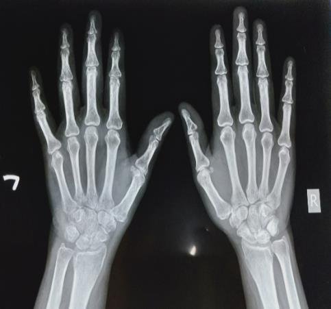
osteopenia (fig.2) is seen in subacute stage and extreme osteopenia/osteoporosis is noted in chronic stage.
confirm whether it is sympathetic mediated pain or not. This is performed usually under fluoroscopy or USG guidance as vital structures are present nearby.
Early recognition is the key of management. Treatment of CRPS requires an individualized
comprehensive rehabilitation protocol on holistic approach to achieve complete recovery and prevent from progressing to irreversible stage of CRPS (Pachydermia). Comprehensive rehabilitation protocol consists of pharmacological therapy (medication), Nonpharmacological therapy (Physiotherapy, occupational therapy etc.) & interventional procedure. Surgery is the treatment of choice when
conservative management fails.
Pharmacological therapy: Following medicines are initially prescribed for management of pain, inflammation or swelling and demineralization:-
Non pharmacological therapy: Apart from medications, role of non-pharmacological therapy is utmost important. Therapeutic exercises like Joint Range of motion exercises, stretching & strengthening
exercises are advised primarily to improve limb mobility. TENS, Paraffin bath & Hyperbaric oxygen therapy have significant role in management of pain, swelling & stiffness. rTMS is suggested in some studies for neuromodulation. Desensitization technique, Hand function training and gait training are important for functional improvement. There are some positive evidences of mirror therapy (fig.5), Qigong, acupuncture for treatment of CRPS.
Interventional procedures: When noninvasive methods are not responsive, interventional procedures are planned. Following regional procedures are strongly recommended by various literatures:
agent and corticosteroid is the treatment of choice. It is a day care procedure usually done under USG or fluoroscopy guidance. Stellate ganglion is located anterior to transverse process of C7 and neck of first rib. Patient is positioned supine with a thin pillow under neck. Head rotated slightly to the opposite side. On fluoroscopic technique, needle is directed to the junction of vertebral body
and transverse process of C6 or C7 between trachea and carotid artery. After bony contact, needle is slightly withdrawn to rest outside of longus coli muscle. Contrast agent is injected to confirm
appropriate needle position and to rule out intravascular or neuraxial injection. After confirmation by both antero-posterior & lateral view, mixture of local anesthetic agent and corticosteroid is
injected. Onset of Horner syndrome (Ptosis, miosis, anhidrosis) with increased temperature (>3F) of affected limb is usually observed in successful block. On USG guided technique, transducer is placed over C6 level. After retracting carotid artery laterally, needle is inserted paratracheally using an in-
plane approach. Following a negative aspiration test for blood or CSF, local anesthetic agent and corticosteroid are injected to subfascial plane of prevertebral fascia.
anesthetic agent and corticosteroid is injected. On USG guided approach, patient is positioned on lateral decubitus with affected limb upwards. Needle is inserted below the lower pole of kidney towards antero-lateral aspect of L2 or L3 vertebral body. After repeated negative aspiration, local anesthetic & corticosteroid are injected on real time visualization. Patient is monitored for 2 hrs and may be discharged after 4 hrs.
Surgery is the treatment of choice in patients refractory to conservative therapy and interventional procedures. Spinal cord stimulation, percutaneous cervicothoracic or lumbar sympathectomy and intrathecal pump of morphine are the effective surgical procedures for CRPS.
Prognosis of CRPS is highly variable. Better outcome is seen in younger persons, children and teenagers. For older people, good outcome may be noticed but some persons experience severe pain and disability despite treatment. Evidences suggest early treatment, particularly rehabilitation, is helpful in limiting
disability.
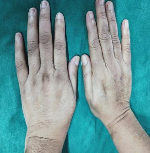
Figure 1: Left upper limb CRPS following supracondylar fracture
Figure 2: X-ray of both hand (AP view) in left upper limb CRPS
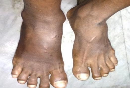
Figure 3: Right lower limb CRPS
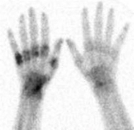
Figure 4: Three-phase bone scintigraphy in left upper limb CRPS
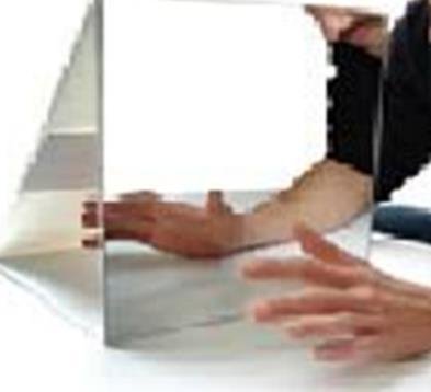
Figure 5: Mirror therapy
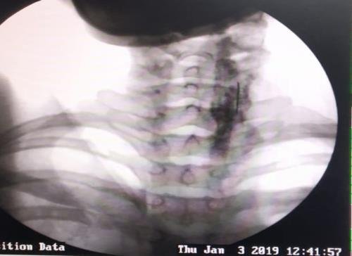
Figure 6: Stellate ganglion block under fluoroscopy guidance
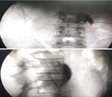
Figure 7: Fluoroscopic guided lumbar sympathetic ganglion block